
Mane Tavadyan1*
1General Medicine, Yerevan State Medical University
*tavman2003@mail.ru
Abstract
The glymphatic system, a glial-dependent cerebrospinal fluid (CSF) transport network, plays a key role in clearing neurotoxic solutes such as amyloid-β and tau. Rodent studies indicate that CSF flows along perivascular spaces and exchanges with interstitial fluid (ISF) to remove metabolic waste at increased rates during sleep. However, most evidence comes from studies in anesthetized animals. In contrast, human data are largely correlative, making it unclear whether glymphatic activity is causative or merely associated with these effects.
This study builds on four hypotheses, slow-wave oscillations drive CSF–ISF exchange, sleep disruption impairs clearance, targeted interventions can enhance glymphatic function and genetic and environmental factors shape vulnerability. This narrative review proposes sleep functions as the primary physiological modulator of glymphatic transport. Integrating anatomical, physiological and molecular evidence, we suggest that disrupted sleep architecture impairs glymphatic dynamics, reducing the clearance of neurotoxic proteins. Conversely, improving sleep quality and modulating key factors, such as vascular pulsatility, aquaporin-4 polarization and lifestyle behaviors, may restore clearance efficiency and offer novel preventive or therapeutic strategies.
Despite progress in glymphatic research, three major gaps persist: reliance on rodent data, lack of standardized human biomarkers and limited understanding of gene–environment interactions. Future research should focus on imaging-based metrics, incorporate genetic profiling into longitudinal studies and develop interventions that enhance slow-wave activity.
In conclusion, sleep emerges not merely as a restorative state but as an active regulator of metabolic clearance, where disruption of either may serve as both a cause and a consequence of neurodegenerative disease.
Introduction
The brain is among the most metabolically active organs in the body (Wang et al. 2011), yet it lacks a classical lymphatic system to clear waste (Liao and Padera 2013). For decades, it remained unclear how neurotoxic byproducts, such as β-amyloid and tau proteins, were removed from the central nervous system (CNS). The discovery of the glymphatic system provided a new and compelling explanation. Emerging evidence highlights sleep as a key regulator of glymphatic activity (Reddy and van der Werf 2020; Ma et al. 2025). During slow-wave sleep, glymphatic transport intensifies, facilitating the efficient removal of neurotoxic solutes (Iliff et al. 2012). Conversely, chronic sleep fragmentation or insufficient sleep impairs this process, potentially accelerating neurodegeneration (Hablitz et al. 2020). Both sleep and glymphatic function decline with age and are disrupted in Alzheimer’s disease (Nedergaard and Goldman 2021). This raises a fundamental question: does impaired sleep cause glymphatic dysfunction and neurodegeneration, or is it a downstream consequence of disease pathology? While research on the glymphatic system has grown rapidly, existing reviews remain fragmented. Most emphasize either the anatomical and physiological basis of glymphatic flow or its clinical implications, but few integrate sleep architecture, molecular genetics, lifestyle influences and translational challenges into a unified framework. To address this gap, the present review is organized around four central hypotheses that collectively capture the most testable mechanisms linking sleep and glymphatic function.
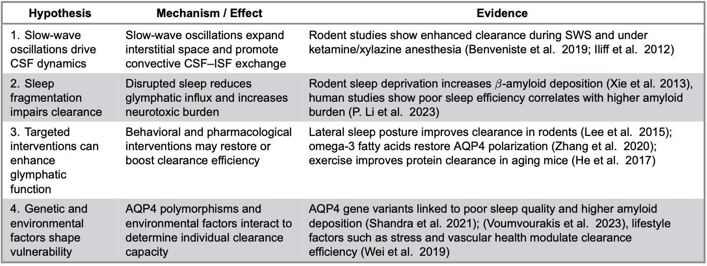
Table 1. Core hypotheses linking sleep and glymphatic function, proposed mechanisms and supporting evidence.
By evaluating the evidence for each of these hypotheses, this review synthesizes current findings on the anatomical and physiological foundations of the glymphatic system, emphasizing its core components and cerebrospinal fluid (CSF) pathways. This review further examines how sleep architecture modulates glymphatic transport and maintains brain homeostasis. Focus is given to both natural and modifiable factors that affect glymphatic efficiency, including vascular pulsatility, aquaporin-4 polarization and lifestyle behaviors such as exercise, stress and sleep posture. Comparison findings from animal and human studies allows for identification of translational challenges and methodological limitations. Integration of molecular genetics and interventional evidence outlines future directions for biomarker validation, imaging advancement and the development of preventive or therapeutic strategies for neurodegenerative disease.
Methodology
This work is a narrative review of the literature on the glymphatic system and its relationship to sleep. Relevant studies were identified through PubMed and Scopus searches. Search terms included combinations of glymphatic system, sleep, CSF clearance, aquaporin-4, slow-wave activity, sleep fragmentation and neurodegeneration. Additional references were located by screening the bibliographies of key publications.
The review focused primarily on articles published from 2012–2025, reflecting the period since the glymphatic system was first described. However, earlier foundational works were also included when they provided essential historical or physiological context.
Only full-text, peer-reviewed articles in English were considered. Articles available only in abstract form, non-peer-reviewed preprints, case reports, commentaries or opinion pieces without empirical data were excluded. Duplicate records retrieved across databases were also removed. Studies were categorized by species (rodent/human), method (e.g., imaging, tracer infusion, biomarker analysis) and strength of inference (mechanistic vs. correlational) to allow for comparative evaluation across the four hypotheses. As this is a narrative review, the PRISMA guidelines were not applicable.
Anatomical and Physiological Foundations
Cerebrospinal Fluid Pathways and Brain Fluid Compartments
Unlike peripheral tissues, the CNS lacks conventional lymphatic vessels (Liao and Padera 2013). Instead, the CNS relies on CSF and unique brain fluid compartments to maintain homeostasis. These compartments include intracellular fluid (60–68%), interstitial or extracellular fluid (12–20%), blood (10%) and CSF (10%) (Damkier et al. 2013). CSF is primarily produced by the choroid plexus located within the brain’s ventricles. It circulates through the ventricular system, flowing from the lateral ventricles through the foramen of Monro to the third ventricle and ultimately to the fourth ventricle via the aqueduct of Sylvius. CSF then exits into the subarachnoid space via the foramina of Magendie and Luschka (Damkier et al. 2013). Roughly 500–600mL of CSF is produced daily in humans, refreshing the ~150mL total volume four times per day (Damkier et al. 2013; Jessen et al. 2015). This renewal ensures continuous nutrient delivery and waste removal.
Although the circulation of CSF is well characterized, the exact contribution of CSF turnover to metabolite clearance remains under debate. Some argue that CSF renewal alone cannot explain efficient clearance of β-amyloid and tau (Elbert et al. 2022; Li et al. 2022), which motivated the discovery of a more active mechanism.
The Brain’s Barriers: Blood-Brain and Blood-CSF Interfaces
Fluid flow in the brain is tightly regulated by two crucial barriers: the blood-brain barrier (BBB) and the blood–CSF barrier (shown in Figure 1) (Dotiwala et al. 2023). The BBB is formed by endothelial cells with tight junctions, pericytes and astrocytic endfeet, restricting paracellular transport (Wu et al. 2023). The blood–CSF barrier, located at the choroid plexus, relies on tight junctions between epithelial cells and selectively regulates solute composition in CSF (Jessen et al. 2015). While these barriers protect brain homeostasis, they also complicate therapeutic delivery (Wu et al. 2023; Katz et al. 2025). Thus, understanding how the glymphatic system interfaces with these barriers could unlock drug delivery strategies.
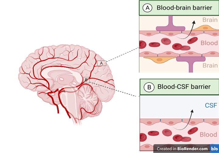
Figure 1. The blood–brain barrier is formed by endothelial cells with tight junctions, pericytes and astrocytic endfeet, whereas the blood–CSF barrier at the choroid plexus relies on epithelial tight junctions to regulate solute composition in CSF. Their interaction with glymphatic pathways highlights the importance of perivascular spaces and astrocytic aquaporin-4 in coordinating CSF–ISF exchange.
Perivascular Spaces and Astrocytic Networks
Cerebral arteries on the cortical surface branch into pial arteries which run along the brain surface within the pia mater and then descend into penetrating arterioles that extend into the brain parenchyma. These penetrating vessels create perivascular (Virchow-Robin) spaces, fluid-filled channels surrounded on the outside by astrocytic endfeet (Jessen et al. 2015). These channels provide conduits for CSF influx into interstitial compartments.
Astrocytes are essential regulators of brain fluid movement due to their high expression of aquaporin-4 (AQP4) on endfeet membranes. AQP4 is a water channel protein that facilitates CSF–ISF exchange (Papadopoulos and Verkman 2013). In the brain, AQP4 is predominantly localized to subpial astrocyte processes, forming the glial-limiting membrane that interfaces between the central nervous system (CNS) and CSF. It is also present in perivascular astrocytic endfeet at the CNS–blood interface (Papadopoulos and Verkman 2013). This specific distribution is crucial for enabling bulk fluid movement between influx and efflux pathways. Notably, knockout studies have shown that deletion of AQP4 in astrocytes reduces CSF influx and glymphatic clearance by approximately 70%, underscoring its pivotal role in brain fluid transport (Iliff et al. 2012).
The Glymphatic System as a Structured Fluid Transport Network
Discovery and Conceptual Foundation
Although ISF was once thought to move solely by diffusion, tracer studies have shown that perivascular drainage occurs via bulk fluid flow, which is markedly faster than passive diffusion (Plog and Nedergaard 2018).
In 2012, a brain-wide perivascular network supporting CSF–ISF exchange was identified using fluorescent tracers in mice, leading to the introduction of the term “glymphatic system” (Jessen et al. 2015). These tracers revealed rapid CSF influx along paravascular spaces surrounding cortical arteries, followed by interstitial penetration and clearance along perivenous pathways (Jessen et al. 2015). This discovery reframed neurobiology by showing that clearance is not passive but organized and state-dependent. However, direct confirmation of this system in humans remained technically challenging, with most evidence inferred from MRI-based proxies (Boyd et al. 2024; Satpathy et al. 2025).
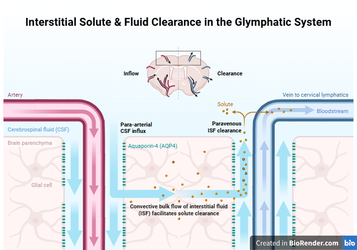
Figure 2. Perivascular (Virchow-Robin) spaces surrounding penetrating arterioles act as pathways for CSF influx into interstitial compartments. Astrocytic endfeet enriched with AQP4 facilitate CSF–ISF exchange, making AQP4 essential for efficient glymphatic clearance.
Core Functions of the Glymphatic System
With its primary function being the pseudo-lymphatic clearance of waste, the glymphatic system supports multiple interconnected functions that are essential to maintaining brain homeostasis (shown in Table 2). Its most well-established role lies in waste clearance, particularly in the removal of β-amyloid, tau and lactate from the brain (Jessen et al. 2015). In addition, it facilitates nutrient and lipid transport, including the movement of apolipoprotein E and lipophilic molecules smaller than 1kDa, which contribute to astrocytic calcium signaling (Rangroo Thrane et al. 2013). The system also supports metabolic homeostasis, as lactate clearance is markedly enhanced during sleep, with experimental suppression of glymphatic activity shown to disrupt this balance (Lundgaard et al. 2018). Beyond these roles, cerebrospinal fluid-mediated solute transport has been suggested to hold therapeutic potential for improving CNS drug delivery (Lohela et al. 2022; Katz et al. 2025). Finally, the system may contribute to immune surveillance, given evidence that cerebrospinal fluid signaling interfaces with peripheral immunity (Nycz and Mandera 2021). Importantly, while the waste-clearing function is strongly supported in animal studies, more speculative roles such as drug delivery and immune surveillance remain largely hypothetical in humans.
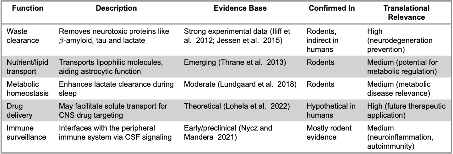
Table 2. Proposed functions of the glymphatic system, their supporting evidence, experimental confirmation and translational relevance.
Sleep as a Primary Physiological Driver of Glymphatic Function
Sleep and the Glymphatic System
Sleep is a fundamental physiological process that plays a critical role in maintaining brain homeostasis. Despite reducing behavioral activity and increasing vulnerability, it has been evolutionarily conserved across species, highlighting its essential restorative functions (Zielinski et al. 2016). It is a highly organized neurophysiological process composed of non-rapid eye movement (NREM) and rapid eye movement stages, each marked by distinct oscillations and autonomic profiles. During NREM, especially slow-wave sleep (SWS), synchronized cortical delta waves, reduced sympathetic tone and increased parasympathetic activity create an optimal environment for metabolic recovery (Reddy and van der Werf 2020; Deng et al. 2024). Building upon its structural and functional framework, the glymphatic system operates as a dynamic, state-dependent clearance network whose efficiency is profoundly influenced by sleep architecture.
Although overall metabolic rate declines modestly (~20%) during sleep, the expansion of interstitial space during SWS markedly enhances CSF–ISF exchange (Jessen et al. 2015; DiNuzzo and Nedergaard 2017). Glymphatic activity peaks in this phase, when reduced neuronal firing and vascular tone facilitate convective solute clearance (Benveniste et al. 2019; Jiang-Xie et al. 2024).
However, translating these observations from animal models to humans presents several challenges. In rodent studies, a 9% increase in interstitial volume has been documented during sleep (Benveniste et al. 2019), but it remains unclear whether similar proportional changes occur in the human brain. Compared to rodents, humans have larger brains, slower CSF turnover and less consolidated SWS, particularly with aging (Liang et al. 2022; Miao et al. 2024). Human studies have primarily relied on indirect markers, such as EEG-CSF coupling, amyloid PET imaging or diffusion tensor imaging along the perivascular space (DTI-ALPS), which are largely correlational and lack mechanistic specificity. Furthermore, the extent to which sleep facilitates glymphatic clearance in humans remains an open question.
The clinical implications of impaired sleep are particularly relevant in the context of neurodegenerative disease. Sleep disturbances, including insomnia, obstructive sleep apnea and circadian rhythm disorders, are associated with decreased glymphatic efficiency and higher risk of cognitive decline (Baranwal et al. 2023). In Alzheimer’s disease (AD), sleep disruption often precedes the onset of cognitive symptoms, suggesting a role in early pathogenesis. Individuals with elevated amyloid frequently exhibit reduced sleep efficiency and increased daytime napping due to impaired circadian regulation and melatonin signaling (Anghel et al. 2023; Li et al. 2023). Despite these associations, sleep disorders are clinically diagnosed in only about one-third of AD patients, suggesting under-recognition or misattribution to normal aging (Reddy and van der Werf 2020). The bidirectional relationship between sleep disruption and amyloid burden complicates therapeutic approaches by raising the question of whether interventions should target sleep to enhance clearance or reduce amyloid to improve sleep quality.
In addition to sleep architecture and duration, sleep posture may also modulate glymphatic activity. Animal models indicate that the right lateral position enhances glymphatic flow, while prolonged time in the supine position is associated with reduced clearance efficiency and greater dementia risk (Lee et al. 2015; Levendowski 2019). These findings suggest that posture during sleep may represent a simple yet modifiable factor influencing brain health.
Finally, genetic factors also shape glymphatic function. AQP4 plays a central role in mediating CSF–ISF exchange (Zhang et al. 2020). Specific single-nucleotide polymorphisms in the AQP4 gene have been associated with reduced sleep quality and increased amyloid accumulation, indicating a potential genetic vulnerability to glymphatic dysfunction (Chandra et al. 2021; Voumvourakis et al. 2023). These gene–environment interactions may partially explain individual differences in susceptibility to neurodegeneration and highlight the need for personalized approaches in glymphatic research.
Multimodal Drivers of Glymphatic Transport
Natural Drivers of Glymphatic Transport
Glymphatic transport depends on a set of physiological drivers that regulate CSF influx and interstitial solute clearance. Among these, arterial pulsation has received the greatest attention. Rodent experiments demonstrate that reducing pulsatility impairs CSF influx (Iliff et al. 2012; Han et al. 2024). Yet, extrapolation to humans remains problematic: arterial stiffening in aging and hypertension alters pulsatile dynamics in ways that animal models may not capture (Mitchell 2021). While Iliff et al. (2012) emphasized the role of arterial smooth muscle contractions, Mestre et al. (2018) proposed that perivascular pumping is augmented by astrocytic and CSF pressure gradients, suggesting a multifactorial mechanism rather than purely vascular mechanism. Whether human glymphatic decline is primarily vascular or instead reflects broader changes in CSF production and astrocytic function remains unresolved. More refined imaging tools that can quantify arterial–CSF coupling in vivo are needed. CSF production, largely determined by choroid plexus function, represents another potential regulator (Damkier et al. 2013). Animal studies suggest that higher CSF production enhances convective flow, yet human studies rarely distinguish whether changes in clearance reflect production, efflux or both (Margetis and Baker 2025). Liu et al. (2020) reported that anesthetic modulation alters CSF secretion patterns without proportional increases in solute clearance, contrasting with earlier work by Damkier et al. (2013), who linked CSF production directly to convective transport efficiency. This inconsistency complicates therapeutic strategies aimed at stimulating production alone.
Respiratory dynamics add further complexity. Slow, deep breathing has been shown to augment CSF movement in MRI and physiological studies (Klose et al. 2000). However, clinical relevance is still unclear, particularly in populations with sleep-disordered breathing. Recent human MRI data suggest that respiration-induced pressure waves primarily influence cerebrospinal fluid displacement within the spinal subarachnoid space, raising questions about their relative contribution to cortical clearance compared with arterial pulsatility (Dreha-Kulaczewski et al. 2018). Whether respiratory-driven CSF movement compensates for vascular decline or instead exacerbates clearance failure during fragmented sleep remains unknown.
Finally, the state of arousal profoundly modulates glymphatic flow. During wakefulness, high norepinephrine (NE) tone constricts interstitial spaces, restricting fluid exchange, whereas slow-wave sleep reduces NE levels and expands interstitial volume (Jessen et al. 2015). This mechanistic link between sleep and clearance is compelling, but current human data are largely correlative, relying on EEG-CSF coupling studies rather than direct fluid measurements. Moreover, the threshold at which sleep fragmentation becomes biologically disruptive remains poorly defined.
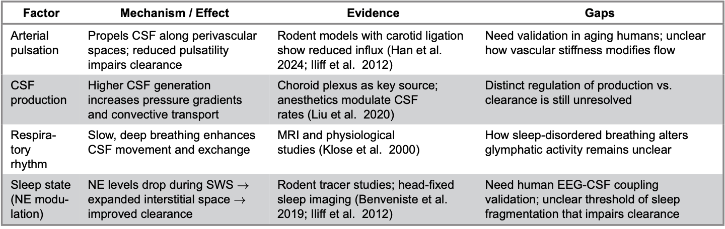
Table 3. Natural drivers of the glymphatic system, their mechanisms and research gaps.
Disruption and Modulation of Glymphatic Function in Aging and Disease
Disruption and Enhancement of Glymphatic Function
Beyond natural physiological drivers, glymphatic activity can be influenced by lifestyle, environmental and genetic factors that either enhance or impair clearance efficiency. The third and fourth hypotheses propose that targeted interventions can augment glymphatic function, while genetic and environmental variability determine individual vulnerability. Aging consistently reduces clearance by ~40%, associated with reduced CSF production, arterial stiffening and loss of AQP4 polarization (Fleischman et al. 2012; Silva et al. 2021). Yet, it remains unclear whether this decline is intrinsic to aging or secondary to vascular comorbidities.
Chronic stress represents a parallel pathway of disruption, elevating norepinephrine and impairing AQP4 expression (Wei et al. 2019). Interestingly, stress-induced NE elevation mimics the arousal-associated glymphatic suppression described by Jessen et al. (2015), implying that stress may phenocopy wakefulness-related glymphatic inhibition. Despite this phenomenon being conceptually consistent with sleep research, direct human evidence is lacking. Type 2 diabetes produces an even more complex phenotype: enhanced influx but impaired efflux, leading to solute buildup (Jiang et al. 2017). This pattern suggests glymphatic failure cannot be reduced to “flow rate” alone; exit routes such as meningeal lymphatics may be equally critical (Gallina et al. 2021).
Meanwhile, lifestyle factors can mitigate or exacerbate these effects. Physical exercise enhances glymphatic clearance, particularly in aging brains (He et al. 2017). Although no significant changes were observed in blood-brain barrier permeability, exercise improved interstitial fluid drainage, AQP4 distribution and synaptic plasticity. The authors noted that enriched cage environments may have contributed to the observed neuroprotective effects, suggesting a combined role for physical and environmental stimulation (He et al. 2017).
Omega-3 fatty acids play a protective role in maintaining glymphatic function, particularly following traumatic brain injury (TBI) (Zhang et al. 2020). In animal models, Omega-3 supplementation restored AQP4 expression and polarization, essential for efficient CSF–ISF exchange (Zhang et al. 2020). Epidemiological data also associate high dietary Omega-3 intake with a lower risk of dementia in aging populations, likely through sustained support of astrocytic and vascular function (Wei et al. 2023).
Alcohol exhibits a clearly dose-dependent pattern—low levels enhance glymphatic clearance, whereas chronic or heavy exposure impairs it (Lundgaard et al. 2018). While these findings support a hormetic model of glymphatic regulation, later imaging data (Drouka et al. 2025) argue that even moderate intake may reduce AQP4 polarization in predisposed individuals, challenging the idea of a universally safe threshold. However, clinical evidence remains inconsistent. Several cross-sectional and cohort studies have produced conflicting results regarding the association between alcohol consumption and Alzheimer’s-related biomarkers. A large-scale study involving 806 participants reported that moderate alcohol use (≥1 time/week) correlated with higher cerebrospinal fluid (CSF) β-amyloid and tau ratios, suggesting a potentially detrimental influence compared with light drinking (≤1 time/week) (Drouka et al. 2025). Conversely, smaller cohorts have shown the opposite trend, indicating a possible protective association between moderate consumption and reduced amyloid-beta (Aβ) burden in cognitively healthy individuals (Vassilaki et al. 2018). Still, some studies found no significant relationship between alcohol intake and CSF Aβ or tau deposition (Koch et al. 2020).
A summary of the natural drivers and modifiable factors affecting glymphatic function, along with their proposed mechanisms and impact, is provided in Table 4.
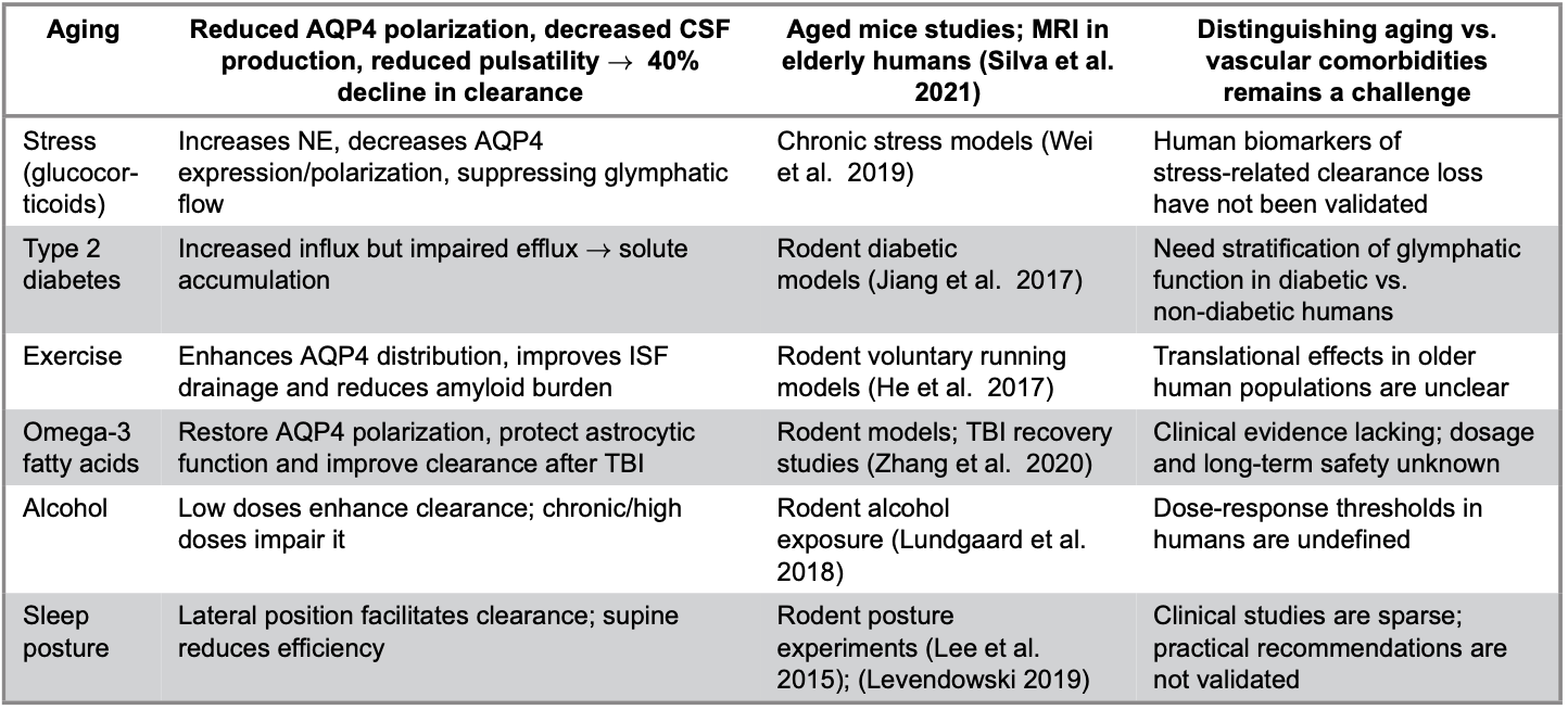
Table 4. Modifiable factors of the glymphatic flow.
Discussion
This review proposes that sleep functions as the principal physiological modulator of glymphatic clearance, linking neurophysiological rhythms to brain waste removal and long-term neurological resilience. Building on four hypotheses, (1) slow-wave oscillations drive cerebrospinal–interstitial fluids exchange, (2) sleep disruption impairs clearance, (3) targeted interventions can enhance glymphatic function and (4) genetic and environmental factors shape vulnerability. The analysis synthesizes evidence across molecular imaging and behavioral domains to elucidate the glymphatic axis as a bidirectional and modifiable system central to neurodegenerative disease risk and prevention.
Taken together, the collective evidence suggests that sleep-dependent modulation of glymphatic activity contributes to long-term neurological resilience, particularly in the context of aging and neurodegenerative risk (Iliff et al. 2012; Benveniste et al. 2019; Han et al. 2024). More importantly, the evidence supports a bidirectional relationship in which sleep both influences and is influenced by clearance efficiency, reframing the sleep–glymphatic axis as a dynamic and context-dependent process rather than a linear causal pathway.
However, the lack of uniform support for the sleep–glymphatic paradigm carries significant implications for how glymphatic biology should be interpreted and applied. The modest diurnal changes observed in diffusion MRI and the temporal dissociation between sleep disruption and amyloid deposition in PET studies suggest that glymphatic transport alone is unlikely to be a dominant or sufficient driver of solute clearance in the human brain (Ringstad 2024). Rather than invalidating the glymphatic model, findings indicate that its contribution may be conditional, state-dependent and constrained by parallel clearance mechanisms, particularly vascular and meningeal lymphatic pathways (Jessen et al. 2015; Boyd et al. 2024; Satpathy et al. 2025). From an applied perspective, this redundancy implies that interventions targeting glymphatic flow in isolation are unlikely to yield robust or universal clinical benefits. The persistence of solute clearance in AQP4-deficient models and the apparent dominance of diffusion and vascular transport in humans suggest that compensatory mechanisms may buffer partial dysfunction (Smith et al. 2017). Consequently, therapeutic strategies aimed solely at enhancing sleep quality or astrocytic water transport may be insufficient unless accompanied by interventions that also improve vascular compliance, cerebrospinal fluid turnover or lymphatic drainage (Ray et al. 2021). This may explain why lifestyle and behavioral interventions that enhance glymphatic markers in rodents fail to translate consistently into measurable biomarker or cognitive benefits in human studies.
Lifestyle evidence is equally mixed. Mild alcohol exposure enhances glymphatic clearance in rodents (Lundgaard et al. 2018), yet human data are inconsistent. Some studies associate moderate consumption with increased amyloid and tau levels (Helseth et al. 2023), whereas others report neutral or protective outcomes (Vassilaki et al. 2018; Koch et al. 2020; Drouka et al. 2025). This discrepancy implies a nonlinear, context-dependent relationship. Similarly, while exercise and omega-3 fatty acids promote clearance and astrocyte function in animal models, their benefits in humans remain uncertain due to age-related and metabolic confounders (He et al. 2017; Zhang et al. 2020).
These inconsistencies highlight several unresolved scientific gaps that limit translational progress. First, causal relationships between sleep disruption, glymphatic dysfunction and neurodegenerative pathology remain unclear in humans. Most available evidence is correlational, and longitudinal PET studies suggest that amyloid accumulation may precede sleep impairment in certain populations, supporting a bidirectional feedback loop rather than a unidirectional causal pathway. Species differences further complicate interpretation: human brains exhibit slower cerebrospinal fluid production and less consolidated slow-wave activity than rodents, which may result in fundamentally different clearance kinetics (Simon et al. 2016; Deng et al. 2024). Consequently, while rodent studies illuminate mechanistic principles, direct extrapolation risks oversimplification (key translational constraints are summarized in Table 5).
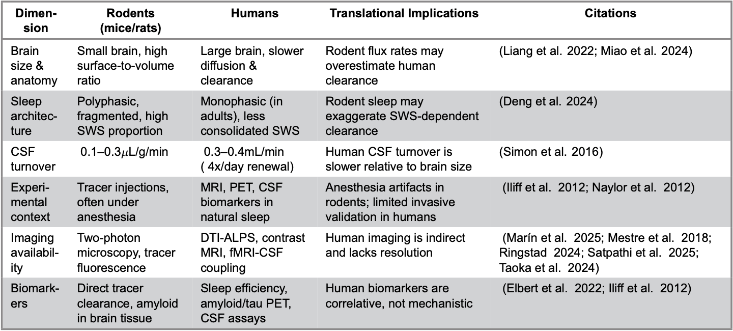
Table 5. Differences between rodent and human brain structure, their implications on the glymphatic flow
Second, technical barriers have limited our ability to confirm glymphatic function in humans. Most evidence derives from advanced MRI techniques, such as DTI-ALPS indices, intrathecal contrast-enhanced imaging and fMRI-CSF coupling (Ringstad 2024; Taoka et al. 2024; Marín et al. 2025). These tools are either invasive, indirect or prone to artifacts, making cross-study comparisons unreliable. Correlations between EEG slow-wave activity and CSF pulsatility are compelling (Reddy and van der Werf 2020), but they remain associative rather than mechanistic. Progress will require standardized imaging protocols and multimodal integration, linking MRI with electrophysiology, PET tracers and CSF biomarkers, to capture both spatial and temporal dynamics of clearance.
Third, biomarker development remains incomplete. Lactate, amyloid, tau and sleep efficiency have all been proposed as markers of glymphatic activity, yet none are specific (Naylor et al. 2012; Elbert et al. 2022). Multimodal biomarker profiles that integrate molecular measures with imaging readouts may offer greater sensitivity, but causality remains unresolved. Evidence that amyloid itself disrupts AQP4 localization and vascular pulsatility supports a bidirectional loop in which impaired clearance and pathology reinforce one another (Iliff et al. 2012; He et al. 2017). Longitudinal human studies integrating sleep architecture, CSF dynamics and molecular biomarkers are therefore essential to disentangle cause from consequence.
Genetic variation adds a further, underexplored dimension. Polymorphisms in AQP4, APOE and vascular-regulatory genes have been linked to differences in clearance efficiency, sleep quality and amyloid burden (Chandra et al. 2021; Voumvourakis et al. 2023). However, these findings remain fragmented and largely absent from intervention trials. Future research should take three steps: (1) embed genotyping into longitudinal cohorts to map genotype–phenotype–outcome relationships; (2) stratify interventional studies to test whether specific variants (e.g., AQP4, APOE4) modulate treatment responsiveness; and (3) adopt multi-omic strategies to reveal how genetic variants influence astrocytic polarization, vascular pulsatility or circadian coupling. Such work could pave the way toward personalized glymphatic medicine, where prevention and therapy are guided by individual clearance profiles.
Lifestyle and behavioral factors, though often highlighted in preclinical work, remain insufficiently integrated into translational studies. Exercise, omega-3 supplementation, sleep posture and even moderate alcohol consumption have been shown to enhance glymphatic transport in rodents, while chronic stress, diabetes and heavy alcohol exposure impair it (Lee et al. 2015; He et al. 2017; Jiang et al. 2017; Levendowski 2019; Wei et al. 2019; Zhang et al. 2020). Yet these findings are rarely validated in humans where confounding vascular and metabolic factors dominate. Determining whether lifestyle modifiers act directly on glymphatic pathways or indirectly through systemic mechanisms such as vascular compliance, inflammation or circadian alignment represents a key priority for future interventional studies.
Finally, enthusiasm for glymphatic biology should not obscure the broader clearance landscape. The glymphatic pathway interacts with meningeal lymphatics, the blood–brain barrier and immune surveillance systems (Hershenhouse et al. 2019; Licastro et al. 2024). Overemphasis on a single pathway risks oversimplification, particularly since compensatory clearance routes may buffer dysfunction under certain conditions. Integrative models, not isolated mechanisms, are essential for accurately situating glymphatic transport within the broader physiology of brain waste management.
Future research should prioritize three major goals. First, there is a need for methodological refinement. Standardized, noninvasive imaging biomarkers such as DTI-ALPS, dynamic PET–CSF coupling and multimodal EEG–fMRI should be developed to quantify glymphatic function in humans with improved temporal and spatial resolution. Second, causal relationships between impaired sleep drivers and the glymphatic clearance should be established. Longitudinal and interventional studies should determine whether impaired sleep drives reduce clearance or instead emerge as a consequence of it, ideally by manipulating slow-wave activity or noradrenergic signaling. Third, genetic and personalized factors should be integrated into study designs. Stratifying participants by AQP4 genotypes will allow researchers to assess individual vulnerability and differential responses to sleep-based or lifestyle interventions. Expansion of these approaches into nonhuman primate models and aging human cohorts will strengthen translational relevance and clinical applicability.
These conflicting findings highlight that sleep is a facilitator, but not the sole determinant of glymphatic clearance. Glymphatic dysfunction likely acts synergistically with vascular aging, inflammation and genetic predisposition to accelerate neurodegeneration.
Clinically, this framework connects sleep quality to cerebrovascular health and cognitive resilience, emphasizing preventive approaches that include slow-wave optimization, vascular maintenance and circadian alignment. Conceptually, it redefines sleep from a passive restorative phase to an active regulator of cerebral fluid homeostasis.
Conclusion
The past decade has redefined brain homeostasis by identifying the glymphatic system as a key mechanism linking sleep to metabolic waste clearance and long-term neurological health. While evidence strongly supports sleep as a driver of CSF–ISF exchange and neurotoxic solute removal, translation to humans remains incomplete. Three gaps remain unresolved. First, methodological barriers, including the lack of standardized, non-invasive biomarkers of glymphatic activity, limit clinical validation. Second, the role of impaired clearance in neurodegeneration is likely bidirectional, functioning both as a driver and a consequence of disease. Third, the system is often studied in isolation, despite clear interactions with meningeal lymphatics, vascular pulsatility and immune surveillance. Overlooking these interdependencies risks oversimplification and may obscure compensatory pathways.
Future research should prioritize four directions: (1) validation of human biomarkers that reliably reflect glymphatic function; (2) longitudinal studies linking sleep architecture, CSF dynamics and cognition; (3) interventional trials testing whether enhancing slow-wave activity or lifestyle changes can improve clearance; and (4) integration of genetic and multi-omic data to develop personalized strategies. Ultimately, addressing these challenges will determine whether targeting the glymphatic system can evolve from an experimental concept into a clinical tool for promoting brain resilience and preventing neurodegenerative disease.
Conflict of Interest
The author declares no conflicts of interest.
Acknowledgments
I would like to express my sincere gratitude to Dr. Sona Galstyan for her guidance and support throughout the writing process of this review. Her insights and encouragement significantly contributed to the clarity and direction of the final manuscript.
References
Anghel, L.C., Ciubară, A., Nechita, A., Nechita, L., Manole, C., Baroiu, L., Ciubară, A.B. and Mușat, C.L. (2023) ‘Sleep disorders associated with neurodegenerative diseases’, Diagnostics, 13(18), 2898. https://doi.org/10.3390/diagnostics13182898
Baranwal, N., Yu, P.K. and Siegel, N.S. (2023) ‘Sleep physiology, pathophysiology, and sleep hygiene’, Progress in Cardiovascular Diseases, 77, 59–69. https://doi.org/10.1016/j.pcad.2023.02.005
Benveniste, H., Liu, X., Koundal, S., Sanggaard, S., Lee, H. and Wardlaw, J. (2019) ‘The glymphatic system and waste clearance with brain aging: A review’, Gerontology, 65(2), 106–119. https://doi.org/10.1159/000490349
Boyd, E.D., Kaur, J., Ding, G., Chopp, M. and Jiang, Q. (2024) ‘Clinical magnetic resonance imaging evaluation of glymphatic function’, NMR in Biomedicine, 37(8), e5132. https://doi.org/10.1002/nbm.5132
Chandra, A., Farrell, C., Wilson, H., Dervenoulas, G., De Natale, E.R., Politis, M. and Alzheimer’s Disease Neuroimaging Initiative (2021) ‘Aquaporin-4 polymorphisms predict amyloid burden and clinical outcome in the Alzheimer’s disease spectrum’, Neurobiology of Aging, 97, 1–9. https://doi.org/10.1016/j.neurobiolaging.2020.06.007
Damkier, H.H., Brown, P.D. and Praetorius, J. (2013) ‘Cerebrospinal fluid secretion by the choroid plexus’, Physiological Reviews, 93(4), 1847–1892. https://doi.org/10.1152/physrev.00004.2013
DiNuzzo, M. and Nedergaard, M. (2017) ‘Brain energetics during the sleep–wake cycle’, Current Opinion in Neurobiology, 47, 65–72. https://doi.org/10.1016/j.conb.2017.09.010
Dreha-Kulaczewski, S., Konopka, M., Joseph, A.A., Kollmeier, J., Merboldt, K.D., Ludwig, H.C., Gärtner, J. and Frahm, J. (2018) ‘Respiration and the watershed of spinal CSF flow in humans’, Scientific Reports, 8(1), 5594. https://doi.org/10.1038/s41598-018-23908-z
Drouka, A., Ntetsika, K.D., Brikou, D., Mamalaki, E., Ntanasi, E., Chatzipanagiotou, S., Gu, Y., Scarmeas, N. and Yannakoulia, M. (2025) ‘Associations of moderate alcohol intake with cerebrospinal fluid biomarkers of Alzheimer’s disease: Data from the ALBION study’, European Journal of Nutrition, 64(3), 142. https://doi.org/10.1007/s00394-025-03651-8
Dotiwala, A.K., McCausland, C. and Samra, N.S. (2023) ‘Anatomy, head and neck: Blood–brain barrier’, in StatPearls [Internet]. Treasure Island, FL: StatPearls Publishing. Available at: https://www.ncbi.nlm.nih.gov/books/NBK519556/
Elbert, D.L., Patterson, B.W., Lucey, B.P., Benzinger, T.L.S. and Bateman, R.J. (2022) ‘Importance of CSF-based Aβ clearance with age in humans increases with declining efficacy of blood–brain barrier/proteolytic pathways’, Communications Biology, 5(1), 98. https://doi.org/10.1038/s42003-022-03037-0
Fleischman, D., Berdahl, J.P., Zaydlarova, J., Stinnett, S., Fautsch, M.P. and Allingham, R.R. (2012) ‘Cerebrospinal fluid pressure decreases with older age’, PLoS ONE, 7(12), e52664. https://doi.org/10.1371/journal.pone.0052664
Gallina, P., Nicoletti, C., Scollato, A. and Lolli, F. (2021) ‘The “glymphatic-lymphatic system pathology” and a new categorization of neurodegenerative disorders’, Frontiers in Neuroscience, 15, 669681. https://doi.org/10.3389/fnins.2021.669681
Hablitz, L.M., Plá, V., Giannetto, M., Vinitsky, H.S., Stæger, F.F., Metcalfe, T., Nguyen, R., Benrais, A. and Nedergaard, M. (2020) ‘Circadian control of brain glymphatic and lymphatic fluid flow’, Nature Communications, 11(1), 4411. https://doi.org/10.1038/s41467-020-18115-2
Han, G., Jiao, B., Zhang, Y., Wang, Z., Liang, C., Li, Y., Hsu, Y.C. and Bai, R. (2024) ‘Arterial pulsation dependence of perivascular cerebrospinal fluid flow measured by dynamic diffusion tensor imaging in the human brain’, NeuroImage, 297, 120653. https://doi.org/10.1016/j.neuroimage.2024.120653
He, X.F., Liu, D.X., Zhang, Q., Liang, F.Y., Dai, G.Y., Zeng, J.S., Pei, Z., Xu, G.Q. and Lan, Y. (2017) ‘Voluntary exercise promotes glymphatic clearance of amyloid beta and reduces the activation of astrocytes and microglia in aged mice’, Frontiers in Molecular Neuroscience, 10, 144. https://doi.org/10.3389/fnmol.2017.00144
Hershenhouse, K.S., Shauly, O., Gould, D.J. and Patel, K.M. (2019) ‘Meningeal lymphatics: A review and future directions from a clinical perspective’, Neuroscience Insights, 14, 1179069519889027. https://doi.org/10.1177/1179069519889027
Iliff, J.J., Wang, M., Liao, Y., Plogg, B.A., Peng, W., Gundersen, G.A., Benveniste, H., Vates, G.E., Deane, R., Goldman, S.A., Nagelhus, E.A. and Nedergaard, M. (2012) ‘A paravascular pathway facilitates CSF flow through the brain parenchyma and the clearance of interstitial solutes, including amyloid β’, Science Translational Medicine, 4(147), 147ra111. https://doi.org/10.1126/scitranslmed.3003748
Jessen, N.A., Munk, A.S., Lundgaard, I. and Nedergaard, M. (2015) ‘The glymphatic system: A beginner’s guide’, Neurochemical Research, 40(12), 2583–2599. https://doi.org/10.1007/s11064-015-1581-6
Jiang, Q., Zhang, L., Ding, G., Davoodi-Bojd, E., Li, Q., Li, L., Sadry, N., Nedergaard, M., Chopp, M. and Zhang, Z. (2017) ‘Impairment of the glymphatic system after diabetes’, Journal of Cerebral Blood Flow & Metabolism, 37(4), 1326–1337. https://doi.org/10.1177/0271678X16654702
Jiang-Xie, L.F., Drieu, A., Bhasiin, K., Quintero, D., Smirnov, I. and Kipnis, J. (2024) ‘Neuronal dynamics direct cerebrospinal fluid perfusion and brain clearance’, Nature, 627(8002), 157–164. https://doi.org/10.1038/s41586-024-07108-6
Katz, J.S., Slika, H., Sattari, S.A., Malla, A.P., Xia, Y., Antar, A., Ran, K. and Tyler, B. (2025) ‘Overcoming the blood–brain barrier for drug delivery to the brain’, ACS Omega, 10(30), 32544–32563. https://doi.org/10.1021/acsomega.5c00364
Klose, U., Strik, C., Kiefer, C. and Grodd, W. (2000) ‘Detection of a relation between respiration and CSF pulsation with an echoplanar technique’, Journal of Magnetic Resonance Imaging, 11(4), 438–444. https://doi.org/10.1002/(SICI)1522-2586(200004)11:4<438::AID-JMRI12>3.0.CO;2-O
Koch, M., Costanzo, S., Fitzpatrick, A.L., Lopez, O.L., DeKosky, S., Kuller, L.H., Price, J., Mackey, R.H., Jensen, M.K. and Mukamal, K.J. (2020) ‘Alcohol consumption, brain amyloid-β deposition, and brain structural integrity among older adults free of dementia’, Journal of Alzheimer’s Disease, 74(2), 509–519. https://doi.org/10.3233/JAD-190834
Lee, H., Xie, L., Yu, M., Kang, H., Feng, T., Deane, R., Logan, J., Nedergaard, M. and Benveniste, H. (2015) ‘The effect of body posture on brain glymphatic transport’, Journal of Neuroscience, 35(31), 11034–11044. https://doi.org/10.1523/JNEUROSCI.1625-15.2015
Li, P., Gao, L., Yu, L., Zheng, X., Ulsa, M.C., Yang, H.W., Gaba, A., Yaffe, K., Bennett, D.A., Buchman, A.S., Hu, K. and Leng, Y. (2023) ‘Daytime napping and Alzheimer’s dementia: A potential bidirectional relationship’, Alzheimer’s & Dementia, 19(1), 158–168. https://doi.org/10.1002/alz.12636
Li, Y., Rusinek, H., Butler, T., Glodzik, L., Pirraglia, E., Babich, J., Mozley, P.D., Nehmeh, S., Pahlajani, S., Wang, X., Tanzi, E.B., Zhou, L., Strauss, S., Carare, R.O., Theise, N., Okamura, N. and de Leon, M.J. (2022) ‘Decreased CSF clearance and increased brain amyloid in Alzheimer’s disease’, Fluids and Barriers of the CNS, 19(1), 21. https://doi.org/10.1186/s12987-022-00318-y
Liao, S. and Padera, T.P. (2013) ‘Lymphatic function and immune regulation in health and disease’, Lymphatic Research and Biology, 11(3), 136–143. https://doi.org/10.1089/lrb.2013.0012
Liu, G., Mestre, H., Sweeney, A.M., Sun, Q., Weikop, P., Du, T. and Nedergaard, M. (2020) ‘Direct measurement of cerebrospinal fluid production in mice’, Cell Reports, 33(12), 108524. https://doi.org/10.1016/j.celrep.2020.108524
Lohela, T.J., Lilius, T.O. and Nedergaard, M. (2022) ‘The glymphatic system: Implications for drugs for central nervous system diseases’, Nature Reviews Drug Discovery, 21(10), 763–779. https://doi.org/10.1038/s41573-022-00500-9
Lundgaard, I., Wang, W., Eberhardt, A., Vinitsky, H.S., Reeves, B.C., Peng, S., Lou, N., Hussain, R. and Nedergaard, M. (2018) ‘Beneficial effects of low alcohol exposure, but adverse effects of high alcohol intake on glymphatic function’, Scientific Reports, 8(1), 2246. https://doi.org/10.1038/s41598-018-20424-y
Ma, J., Chen, M., Liu, G.H., Gao, M., Chen, N.H., Toh, C.H., Hsu, J.L., Wu, K.Y., Huang, C.M., Lin, C.M., Fang, J.T., Lee, S.H. and Lee, T.M.C. (2025) ‘Effects of sleep on the glymphatic functioning and multimodal human brain network affecting memory in older adults’, Molecular Psychiatry, 30(5), 1717–1729. https://doi.org/10.1038/s41380-024-02778-0
Margetis, K. and Baker, S. (2025) ‘Physiology, cerebral spinal fluid’, in StatPearls [Internet]. Treasure Island, FL: StatPearls Publishing. Available at: https://www.ncbi.nlm.nih.gov/books/NBK519007/
Marín, I., Torres, F., Riveros, R., Oliva, B., Vega, J., Saavedra, C., Rojas, S., González, M., Bennett, C., Cox, P. and Chabert, S. (2025) ‘Evaluation of the DTI-ALPS index as a biomarker of the glymphatic system at 1.5T’, The Open Neuroimaging Journal, 18, e18744400389049. https://doi.org/10.2174/0118744400389049250723091323
Mestre, H., Tithof, J., Du, T., Song, W., Peng, W., Sweeney, A.M., Olveda, G., Thomas, J.H., Nedergaard, M. and Kelley, D.H. (2018) ‘Flow of cerebrospinal fluid is driven by arterial pulsations and is reduced in hypertension’, Nature Communications, 9(1), 4878. https://doi.org/10.1038/s41467-018-07318-3
Mitchell, G.F. (2021) ‘Arterial stiffness in aging: Does it have a place in clinical practice?’, Hypertension, 77(3), 768–780. https://doi.org/10.1161/HYPERTENSIONAHA.120.14515
Naylor, E., Aillon, D.V., Barrett, B.S., Wilson, G.S., Johnson, D.A., Johnson, D.A., Harmon, H.P., Gabbert, S. and Petillo, P.A. (2012) ‘Lactate as a biomarker for sleep’, Sleep, 35(9), 1209–1222. https://doi.org/10.5665/sleep.2072
Nedergaard, M. and Goldman, S.A. (2020) ‘Glymphatic failure as a final common pathway to dementia’, Science, 370(6512), 50–56. https://doi.org/10.1126/science.abb8739
Nycz, B. and Mandera, M. (2021) ‘The features of the glymphatic system’, Autonomic Neuroscience, 232, 102774. https://doi.org/10.1016/j.autneu.2021.102774
Papadopoulos, M.C. and Verkman, A.S. (2013) ‘Aquaporin water channels in the nervous system’, Nature Reviews Neuroscience, 14(4), 265–277. https://doi.org/10.1038/nrn3468
Plog, B.A. and Nedergaard, M. (2018) ‘The glymphatic system in central nervous system health and disease: Past, present, and future’, Annual Review of Pathology, 13, 379–394. https://doi.org/10.1146/annurev-pathol-051217-111018
Rangroo Thrane, V., Thrane, A.S., Plog, B.A., Thiyagarajan, M., Iliff, J.J., Deane, R., Nagelhus, E.A. and Nedergaard, M. (2013) ‘Paravascular microcirculation facilitates rapid lipid transport and astrocyte signaling in the brain’, Scientific Reports, 3, 2582. https://doi.org/10.1038/srep02582
Reddy, O.C. and van der Werf, Y.D. (2020) ‘The sleeping brain: Harnessing the power of the glymphatic system through lifestyle choices’, Brain Sciences, 10(11), 868. https://doi.org/10.3390/brainsci10110868
Ringstad, G. (2024) ‘Glymphatic imaging: A critical look at the DTI-ALPS index’, Neuroradiology, 66(2), 157–160. https://doi.org/10.1007/s00234-023-03270-2
Satpathi, S., Reid, R.I., Przybelski, S.A., Raghavan, S., Cogswell, P.M., Meyer, N.K., Lowe, V.J., Gunter, J.L., Petersen, R.C., Jack, C.R., Graff-Radford, J. and Vemuri, P. (2025) ‘Evaluation and interpretation of DTI-ALPS, a proposed surrogate marker for glymphatic clearance, in a large population-based sample’, Alzheimer’s Research & Therapy, 17(1), 191. https://doi.org/10.1186/s13195-025-01842-3
Silva, I., Silva, J., Ferreira, R. and Trigo, D. (2021) ‘Glymphatic system, AQP4, and their implications in Alzheimer’s disease’, Neurological Research and Practice, 3(1), 5. https://doi.org/10.1186/s42466-021-00102-7
Taoka, T., Ito, R., Nakamichi, R., Nakane, T., Kawai, H. and Naganawa, S. (2024) ‘Diffusion tensor image analysis along the perivascular space (DTI-ALPS): Revisiting the meaning and significance of the method’, Magnetic Resonance in Medical Sciences, 23(3), 268–290. https://doi.org/10.2463/mrms.rev.2023-0175
Vassilaki, M., Aakre, J.A., Syrjanen, J.A., Mielke, M.M., Geda, Y.E., Kremers, W.K., Machulda, M.M., Alhurani, R.E., Staubo, S.C., Knopman, D.S., Petersen, R.C., Lowe, V.J., Jack, C.R. and Roberts, R.O. (2018) ‘Mediterranean diet, its components, and amyloid imaging biomarkers’, Journal of Alzheimer’s Disease, 64(1), 281–290. https://doi.org/10.3233/JAD-171121
Voumvourakis, K.I., Sideri, E., Papadimitropoulos, G.N., Tsantzali, I., Hewlett, P., Kitsos, D., Stefanou, M., Bonakis, A., Giannopoulos, S., Tsivgoulis, G. and Paraskevas, G.P. (2023) ‘The dynamic relationship between the glymphatic system, aging, memory, and sleep’, Biomedicines, 11(8), 2092. https://doi.org/10.3390/biomedicines11082092
Wang, Z., Ying, Z., Bosy-Westphal, A., Zhang, J., Heller, M., Later, W., Heymsfield, S.B. and Müller, M.J. (2011) ‘Evaluation of specific metabolic rates of major organs and tissues: Comparison between men and women’, American Journal of Human Biology, 23(3), 333–338. https://doi.org/10.1002/ajhb.21137
Wei, B.Z., Li, L., Dong, C.W., Tan, C.C., Alzheimer’s Disease Neuroimaging Initiative and Xu, W. (2023) ‘The relationship of omega-3 fatty acids with dementia and cognitive decline: Evidence from prospective cohort studies of supplementation, dietary intake, and blood markers’, The American Journal of Clinical Nutrition, 117(6), 1096–1109. https://doi.org/10.1016/j.ajcnut.2023.04.001
Wei, F., Song, J., Zhang, C., Lin, J., Xue, R., Shan, L.D., Gong, S., Zhang, G.X., Qin, Z.H., Xu, G.Y. and Wang, L.H. (2019) ‘Chronic stress impairs the aquaporin-4-mediated glymphatic transport through glucocorticoid signaling’, Psychopharmacology, 236(4), 1367–1384. https://doi.org/10.1007/s00213-018-5147-6
Wu, D., Chen, Q., Chen, X., Han, F., Chen, Z. and Wang, Y. (2023) ‘The blood–brain barrier: Structure, regulation, and drug delivery’, Signal Transduction and Targeted Therapy, 8(1), 217. https://doi.org/10.1038/s41392-023-01481-w
Zhang, E., Wan, X., Yang, L., Wang, D., Chen, Z., Chen, Y., Liu, M., Zhang, G., Wu, J., Han, H. and Fan, Z. (2020) ‘Omega-3 polyunsaturated fatty acids alleviate traumatic brain injury by regulating the glymphatic pathway in mice’, Frontiers in Neurology, 11, 707. https://doi.org/10.3389/fneur.2020.00707
Zielinski, M.R., McKenna, J.T. and McCarley, R.W. (2016) ‘Functions and mechanisms of sleep’, AIMS Neuroscience, 3(1), 67–104. https://doi.org/10.3934/Neuroscience.2016.1.67