

Updates from JYI
Undergraduate Research Funding Available Learn more here.
Best of JYI 2023-2025 is available now! Read the Special Issue here.
Are you able to speak English and one other language? Consider joining JYI's Translations Department! Learn more here.
Special Collection
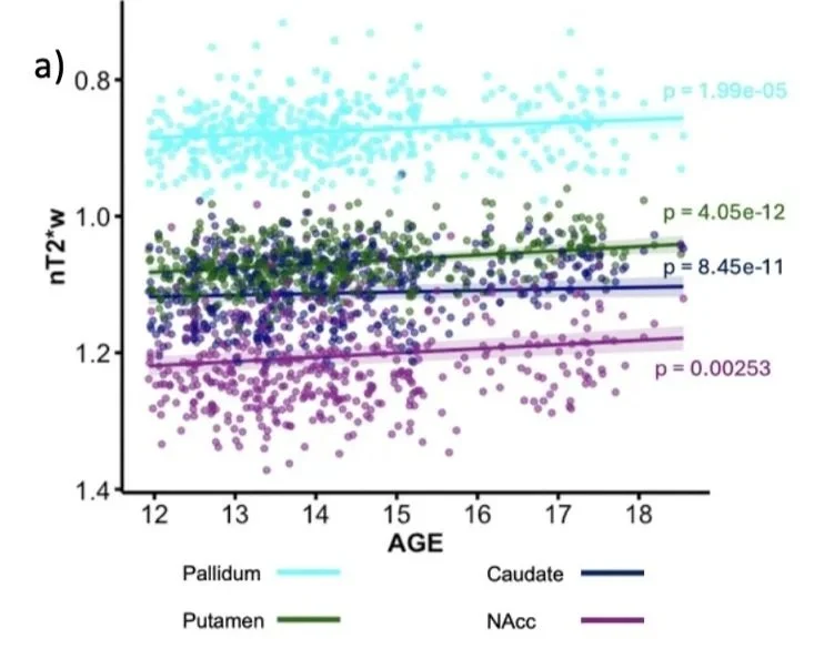
Socioeconomic Disparities in Healthcare Access Special Collection
Accepting on a rolling basis
Featured
2026, May

Featured
The International Undergraduate Research Journal
Interested in submitting your research to the journal?
Follow Us:
Our mission is to improve undergraduate science training by providing innovative, high-quality educational experiences in science writing, publication and the peer-review process.