
Author: Ghatalia Pooja
Institution: Molecular Biology
Date: August 2006
It took only fifty years to realize that it is possible to view bones through flesh using X-rays, that our body can be imaged as thin slices of tissues and that tumors and lesions hidden anywhere in the body can be located. Soon it will be possible to monitor molecular and physiological changes occurring inside the body. Dark-brown iron oxide nanoparticles, about 100,000 times smaller than the diameter of a single hair, can enhance the capabilities of available medical imaging techniques.
These nanoparticles, generally less than 50nm in diameter, are made up of an iron oxide core stabilized by an organic shell. They are called superparamagnetic iron oxide (SPIO) nanoparticles because of their magnetic properties.
An MRI (Magnetic Resonance Imaging) scanner, in a coarse form, consists of a gigantic magnet of 1.5 to 7 Tesla strength (about 600 times stronger than a refrigerator magnet). MR imaging does not involve the use of radiation, which is detrimental to the patient's health. Instead, a magnetic field and radio waves are used to excite all of the hydrogen atoms (called protons) in the body. These protons relax after excitation, and a computer program translates this data into cross-sectional pictures of human tissue.
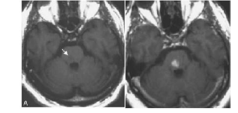
Left - Non-enhanced T1-weighted image brainstem infarct (arrow) Right - Images 48 h (D) following SPIO infusion Image courtesy of: Annals of Biomedical Engineering, Vol. 34, No. 1, January 2006 pp. 2338.
"A key aspect of current imaging is not only the presence of water, but the change in relaxivity of the water molecules near the metal," said Michael Biewer, assistant professor at University of Texas at Dallas. "In other words, the relaxation rate of the excited water molecules is critical for imaging. Normal and diseased tissues can also yield different relaxation rates which can be read by the MRI image."
Dark and light contrast images are created on the computer indicating different relaxation rates. For example, areas of the body with greater water content may appear brighter when compared to some other dense areas. For instance, veins may be brighter than muscles. The greater the magnetic resonance contrast, which helps to create a visual difference between different areas, the better the resolution of the computer image.
Nanoparticles enhance contrast
Before taking the final MRI snapshot, a pre-contrast image is taken. Once the SPIO nanoparticles are injected, a post-contrast image is taken. A clear black-and-white contrast is detected wherever the nanoparticles aggregate in the body.
"If we can direct these nanoparticles to accumulate at a specific location, e.g. a tumor, the nanoparticles will be a vital diagnostic tool leading to an accurate and early detection of diseases," said Chalermchai Khemtong, a postdoctoral fellow at UT Southwestern Medical Center in Texas. "Because of their strong magnetic properties, the SPIO nanoparticles can help us overcome the poor sensitivity of current contrast agents by generating higher contrast between the target and the background." Thus SPIO nanoparticles are a great contrast agent.
Why study the behavior of molecules?
It can help
Early detection of diseases including atherosclerosis (thickening of arterial walls,) thrombosis (formation of clots,) and heart attacks
Understand metastasis of tumors (Metastasis is the migration of tumor from the original tumor site to other tissues or organs)
Track the distribution of cells in the body
These can in turn enable researchers to
Detect solid tumors
Give a personalized treatment depending on the individual's physiological condition, as opposed to a general prescribed treatment
Monitor the effectiveness of the therapeutic treatment
How will you send the nanoparticles into the body?
Magnetic nanoparticles-Lock and Key Mechanism
"If you wish to study, say, the cancer cells, just find out the receptors (proteins) expressed on the surface of cancer cells. If you correlate the receptor to a lock, then all you have to do is tag the SPIO particles with the corresponding key (ligand)," said Khemtong. "When you inject the SPIO particles into the body, after allowing the nanoparticles to bind to the receptors on the cancer cells, you can take MR images and compare images obtained before and after the injection."
To understand this better, imagine a patient undergoing a routine breast cancer scan. To confirm the presence of the cancer cells in her breast, we could inject magnetic SPIO particles into her blood stream. Due to the leaky blood vessels in the tumor, SPIO particles confront the cancer cells and then bind to the breast cancer cells by the "lock and key mechanism". The MRI scanner will produce an excellent contrast image which will help to visualize the presence and location of the tumor.
The biggest challenge in using nanoparticles is targeting them correctly and efficiently to the desired cell or tissue. First, one has to determine the best receptor-ligand combination. Then the receptors to which the magnetic particles are targeted should not only be displayed in excess on the surface of the cells, and should also be unique to the diseased tissue. That is, if the receptors are not specific to the targeted tissue, magnetic particles could bind to undesired tissues of the body and lead to a false diagnosis.
Advantages of using SPIO particles-
Chemical modifications can be made to the SPIO nanoparticles to make them non-toxic, injectable, compatible with the body, and able to be concentrated at a high level in the target tissue or organ.
"SPIO nanoparticles are strongly magnetic so that they are excellent contrast agents for MR imaging. With some advanced development of these agents, a very small amount of these agents will be efficient to produce enough contrast between good and tumorous tissues," said Khemtong. "The surface of the particles can also be chemically modified so they can be friendlier to the body and they can be accumulated in the desired part of the body."
Attempts are made to design magnetic nanoparticles that not only locate tumors and diseases, but also treat them by delivering accurate drug doses. In the near future, we could go for an annual medical examination, in which the doctor would inject SPIO nanoparticles, detect a disease, treat it with nanoparticles, and let us walk out of the room in only a few hours.
References
Akira Ito et al[/]. (2005) Journal of Bioscience and Bioengineering, 100(1) 1-11.
National Cancer Institute, April 2006.
Thorek et al[/]. (2006) Annals of Biomedical Engineering, 34(1) 23-38.
Weissleder. (2006) Journal of American Medical Association, 293(7) 855-862.
*Special thanks to Dr. Khemtong and Dr. Biewer for contributing time for interviews and revisions.