

Author: Frank Adam
Date: August 2005
Chug, chug, chug. An enzyme bustles down a DNA molecule, carefully matching sequences of nucleotides to the new molecule of RNA that it is synthesizing.
Sounds like an excerpt from a high school biology textbook, a simple explanation of the flow of biological information from DNA to RNA, which then codes for a protein. Known as the Central Dogma, this fundamental tenet of biology, developed only within the past 60 years, has truly revolutionized the study of living organisms. But recently a wrinkle seems to have appeared in our understanding of the basic molecules of life and their role in the regulation of genes; a wrinkle that may bring about a second coup in our knowledge of genetics: that of microRNA.
At the forefront of this fledgling field is Gregory Hannon, a scientist at Cold Spring Harbor Laboratory who is attempting to unlock the secrets of gene control. How does a cell know when to turn a gene on or off? How much protein does a gene make? What mechanisms start or stop transcription or translation? These questions are at the core of understanding not only normal cellular function, but also genetic diseases. Hannon has taken what little knowledge we have of microRNA and probed for answers to these big questions in one of the most notable diseases of our time: cancer. His results are intriguing and confirm the upset of the Central Dogma.
The Central Dogma
DNA, deoxyribonucleic acid, carries the information necessary for an organism to grow and develop. This information is housed deep within the nucleus of a cell in genes highly-specific sequences of nucleotides, the building blocks of DNA.
But simply having information does the cell no good. DNA must be able to communicate its instructions out to the body. This is accomplished through mRNA, messenger ribonucleic acid, a single-stranded molecule that bears a close resemblance to the structure of DNA.

Figure 1. A schematic representation of the Central Dogma showing the flow of information from DNA to RNA to protein. Image Courtesy of National Health Museum.
DNA is the foreman of the cell. It constructs mRNA through transcription and sends this molecule out into the cell with orders to create a protein. mRNA does DNA's work: it consults with other molecules in the cell and then translates its genetic information into a functioning protein. Biochemists have termed this whole process "gene expression," since it begins with the genes in DNA (see Figure 1).
Since the inception of modern genetics, this scheme for the flow of information has stood fast. However, Hannon and other scientists are quickly eroding the bedrock of biology, as a result of an amazing discovery . . .
An Amazing Discovery
The Central Dogma defines the role and action of RNA. A few RNAs function solely in translation, but most produce proteins. However, a remarkable discovery was made just over 15 years ago; researchers studying the roundworm Caenorhabditis elegans, an organism frequently used in genetic analysis, found that some RNA wasn't making proteins. Rather, this unique class of RNA was interfering with mRNA to prevent translation, in complete opposition to the Central Dogma. Researchers had stumbled onto a mechanism to control gene expression.
Scientists were once certain that proteins regulated genes. These molecules could bind to DNA and prevent it from transcribing mRNA; or, they could stop mRNA from translating its information into new proteins. In both cases, the proteins ran the show. But it appeared as though something else was also occurring in worms; somehow, special RNA molecules were affecting mRNA and stopping the flow of biological information into proteins.
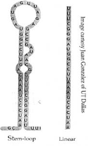
Figure 2. microRNA initially forms the stem-loop structure. Image Courtesy of Juan Gonzales of UT Dallas.
It is no surprise that it took nearly 30 years for scientists to discover this unusual class of RNA, considering that it is over one-thousandth the size of a typical mRNA molecule. Appropriately named microRNA (miRNA), these mini-molecules are encoded by DNA like all RNA. They differ, however, because they do not themselves code for the production of a protein. More exactly, miRNAs do not appear to carry information as mRNA does. For example, in C. elegans the first miRNA molecule discovered, lin-4, suppressed the expression of a specific developmental gene. Other miRNAs were also discovered in plants, fruit flies, mice, and humans, and all seem to play a similar regulatory role.
miRNA is also structurally different than mRNA. miRNA molecules display a truly unique property: folding back on themselves to create a double-stranded structure known as a stem-loop (see Figure 2). This stem-loop shape flags down special enzymes in the nucleus that chop the miRNA molecules into smaller pieces and shuttle them out into the cell. Once here the enzyme Dicer further cuts the miRNA down, bringing it to its tiny working size. Finally, the miRNA binds to a number of proteins collectively known as the RNA-Induced Silencing Complex (RISC) and assumes a linear single-stranded shape. Now the miRNA is ready to go to work in the cell (see Figure 3).
But how exactly do miRNAs carry out their job of interfering with gene expression? mRNA and miRNA both have single-stranded structures in the cell. This property allows miRNA to seek out and bind specific nucleotide sequences in the target mRNA molecule. Here is where researchers are still stumped, though: sometimes miRNA acts like an enzyme itself and chops its mRNA into tiny useless pieces. Other times the miRNA acts like sand in the engine of the cell and simply stalls mRNA, preventing it from translating proteins. In both cases, however, the gene coding for the mRNA is not expressed.
When Regulation Goes Awry
Hannon's interest in miRNA focuses on the chaos that can result from misregulation. A cell is an amazingly complex entity; it carries out precise tasks, interacts with other nearby cells, and responds to both internal and external stimuli in an organized manner. Proper choreography of all aspects of cellular activity hinges on the correct amounts of proteins being present and functioning within a cell at any given time. To achieve this, genes are continually turned on and off in such a way as to affect molecular harmony.
But often a glitch occurs. Perhaps a gene mutates and produces a malfunctioning protein. Maybe a gene is turned on when it should be off. Or perhaps a protein was not broken down at the proper time and now has accumulated within the cell. Unfortunately, circumstances such as these occur often and can precipitate deadly conditions such as cancer.
Cancer represents a loss of a cell's unique identity, which leads to a failure in responding to signals governing growth and differentiation. Thus, cancerous cells proliferate endlessly and eventually invade surrounding tissue. The root of the cell's identity crisis lies in mutations in its DNA.
The Central Dogma states that all of a cell's instructions are contained in DNA. DNA uses this information to produce the proteins that control a cell's actions. A mutation jumbles the genetic data. DNA can no longer create proper proteins, changing how the cell functions. Researchers are interested in how a given mutation affects the constitution of a cell; specifically, what alterations occur in the DNA, RNA, and proteins of a cell?

Figure 3. The schematic shows the major steps in miRNA processing and function. Image courtesy of Charles Mallery University of Miami.
Historically scientists have looked at the total mRNA in a cell using a technique called a microarray. By knowing what mRNA is present, researchers have a good idea of what proteins are being created and to what extent. If, for example, they see that mRNA coding for a growth factor is much more prevalent in a cancer cell, this clues scientists in on the potential cause of that particular type of cancer overproduction of a signal to grow.
Furthermore, cancer biologists are interested in classifying and grouping types of cancer based on similarities in their causes, the cells they affect, and their progressive cycle. Again, by examining what mRNAs are expressed, researchers can find patterns that link forms of cancer together.
Now, however, miRNA has become a hot topic of discussion in this field. Could these little molecular dynamos also play a role in cancer? By examining miRNA expression in a cell can we determine what type of cancer is afflicting a cell? Scientists seem to think so.
Colorful Beads and Oncogenic miRNA
miRNAs were discovered as players in development, a time when cells are continually growing, dividing, and differentiating. Therefore it seems plausible that miRNAs also contribute to growth in cancer.
This is just what Hannon and his colleague Scott Lowe found in B-cell lymphoma, a form of blood cancer. In the June 9 issue of Nature, they reported that a specific cluster of miRNAs, the mir-17-92 cluster, is present in higher concentrations in B-cell lymphoma cells.
To examine more closely mir-17-92's role in cancer, Hannon used the mouse as a model organism. He increased the expression of mir-17-92 in the presence of an oncogene (a gene able to cause cancer) for B-cell lymphoma, c-myc, and infected mice with these cells. His results were striking: the mice with the high concentration of mir-17-92 developed and died from cancer much sooner than mice with normal expression levels. This is substantial evidence in favor of his hypothesis that miRNA can itself function to cause cancer.
In light of his finding, Hannon has proposed accepting a system of nomenclature, developed by researcher Frank Slack, for such oncogenic miRNAs: "oncomiRs."
"We will see lots of different miRNAs touching various aspects of cancer in the future," predicts Hannon. Implementing an organized taxonomy of oncogenic miRNAs now is a key step in recognizing their importance in cancer.
Considering Hannon's findings that miRNA is overabundant in lymphoma cells, it seems likely that examining the expression of miRNA in many different cells may reveal distinct patterns indicative of various forms of cancer.
"Understanding expression profiles of miRNA in cancer is key to identifying tumor types," says Hannon.
In fact, researchers have attempted these experiments, but with only limited success. Unfortunately, due to their small size, miRNA molecules often cause problems for the traditional microarray systems used. However, exciting research in this field has recently come from scientists at MIT. Nobel laureate H. R. Horvitz and fellow researcher Todd Golub have developed a new, highly accurate, and inexpensive system for peering into a cell's collection of miRNA.
Their work, also published in the June 9 issue of Nature, is based on the use of colorful microscopic beads. Tiny plastic beads are given unique colors that correspond to miRNA target molecules present on the beads. miRNA from a cell is then mixed with the beads and binds to its target. The resulting mix is then stained with a fluorescent dye so that researchers can see how much miRNA is actually bound to the beads. By examining the amount of staining for a particular color bead, researchers can catch a glimpse of the miRNA a cell is expressing.
After developing the system, Golub looked at 217 different miRNAs from 334 samples of cells, many of which were cancerous. He noted that miRNA levels were very often reduced in cancerous cells, a finding which might seem to contradict Hannon's work. This is not the case, though; whether increased or decreased, any change in concentration of a regulatory molecule such as miRNA could have dire effects on a cell.
Perhaps even more exciting is the ability of the bead-based detection system to differentiate between various types of cancer. Often the type of cancer afflicting a cell is not easy to determine. This can pose a problem for doctors who need to give very specific treatments for each type of cancer. However, Golub's work may in part solve this medical dilemma, and aid in the diagnosis and treatment of cancer in the future.
"That microRNA profiles have such potential diagnostic utility was a big surprise to us," says Golub. "It's one we're keen to validate in future studies."
Where miRNA is Headed
miRNA, the new kid on the genetic block, has an exciting future in store. Though these molecules were identified in C. elegans as developmental control signals, they seem to be crucial players in proper cellular functioning.
"We will see miRNAs cropping up in many diverse biological processes as research progresses," states Hannon.
Further, Hannon notes that a key aspect for study in years to come is the development of a method to determine what genes are targeted by miRNAs, which will greatly clarify the cellular functions controlled by these molecules.
The study of miRNAs in cancer biology is bound to continue. Hannon's laboratory hopes to better understand the genetic and molecular characteristics of various cancers as they relate to miRNAs. In addition, the potential exists for miRNAs to be a target for cancer therapies.
"There is a chance that nucleic acid antagonists could function in a therapeutic role for cancer patients," says Hannon.
Whatever the aspect, though, research on miRNAs is certain to uncover many exciting and informative duties within the cell for this mini-molecular-dynamo.
Further Reading
Ambros, V et al. (2003). A uniform system for microRNA annotation. RNA. 9:277-9.
Alves, Leonardo. MicroRNA in Systems Biology. Lecture on July 20, 2004.
Cullen, BR. (2004). Transcription and Processing of Human microRNA Precursors. Molecular Cell. 16:861-5.
Golub, TR et al. (2005). microRNA expression profiles classify human cancers. Nature. 435:834-8.
Hannon, GJ et al. (2005). A microRNA polycistron as a potential human oncogene. Nature. 435:828-33.
McManus, MT. (2003). microRNAs and Cancer. Seminars in Cancer Biology. 13:253-8.
Mallory, Charles. RNAi: Interference RNA. University of Miami Department of Biology. Updated: April 18, 2005.
O'Donnell, KA et al. (2005). c-Myc-regulated microRNAs modulate E2F1 expression. Nature. 435:839-43.